


2D Panoramic Dental Radiograph
The panoramic dental radiograph, also known as an orthopantomogram, is a fast and informative diagnostic method that allows the entire dental arch, jaws, temporomandibular joint, and surrounding structures to be seen in a single image. This examination helps to assess dental condition, anomalies in tooth position, cysts, or other pathologies. The orthopantomogram is frequently used in primary diagnosis and treatment planning, particularly prior to orthodontic or surgical procedures.
Our Scanning System
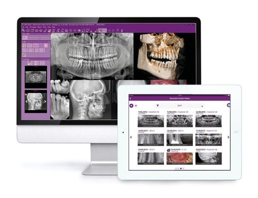
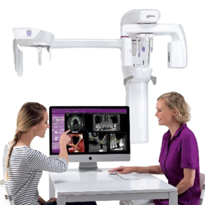
The technology we use allows high-quality panoramic X-ray images to be produced with a minimal radiation dose. We provide convenient online access to completed examinations, and advanced image processing algorithms help dentists to assess patient condition more easily. Unlike general radiology centres, we specialise in dental X-ray examinations, enabling us to maintain the highest professional standards in this field.
Our Scanning System
The technology we use allows high-quality panoramic X-ray images to be produced with a minimal radiation dose. We provide convenient online access to completed examinations, and advanced image processing algorithms help dentists to assess patient condition more easily. Unlike general radiology centres, we specialise in dental X-ray examinations, enabling us to maintain the highest professional standards in this field.
What is a 2D panoramic X-ray?
A 2D panoramic X-ray is a two-dimensional radiographic image showing the patient’s entire dental arch, upper and lower jaws, joints, and sinuses. It is a non-invasive, fast, and effective examination that reveals changes in tooth and bone structure, pathologies, or other important diagnostic findings.
Unlike standard intraoral X-rays, which capture only a small section of the teeth, a panoramic X-ray examination allows the entire oral cavity to be seen in a single image. This examination is indispensable when planning prosthetic treatment, orthodontic therapy, surgical interventions, or general assessment of dental condition.
Why is a panoramic dental X-ray important?
A panoramic X-ray allows the dentist to assess dental condition, bone health, identify orthodontic problems, and detect congenital anomalies. This examination is particularly important when planning dental treatment, orthodontic therapy, or surgical procedures.
Key applications of panoramic X-ray imaging
Orthodontic treatment – leidžia ortodontams įvertinti dantų išsidėstymą ir planuoti gydymo eigą.
Analysis of damaged teeth – padeda nustatyti karieso pažeistus ar negyvus dantis, dantų šaknų pakitimus.
Paranasal sinuses assessment – provides information on the condition of the maxillary sinuses, possible inflammation, or pathology.
Temporomandibular joint (TMJ) disorders sąnario (TMJ) problemos – leidžia įvertinti sąnarių būklę ir galimus pakitimus
Oral pathologies – helps identify bone or soft-tissue pathologies that may be invisible on standard dental X-rays.
Why is a panoramic X-ray sometimes insufficient?
Although a panoramic X-ray examination provides a great deal of valuable information, it does have certain limitations. As it is a two-dimensional image, some details may overlap or be less clearly visible. For example, where a very precise assessment of tooth roots, nerve canals, or bone density is required, cone beam computed tomography (CBCT) may be recommended, as it provides a three-dimensional view.
Panoramic X-ray as a “marketing tool”?
Panoramic X-ray images not only help the clinician to diagnose and plan treatment, but also serve as an excellent visual aid for the patient. Clear and easy-to-understand images allow patients to better appreciate the condition of their teeth, discuss the proposed treatment, and feel confident that they are receiving the best possible care.